What is Wilms tumour?
Wilms tumour starts in immature kidney cells. A cancerous (malignant) tumour is a group of cancer cells that can grow into nearby tissue and destroy it. The tumour can also spread (metastasize) to other parts of the body. Wilms tumour is also called nephroblastoma.
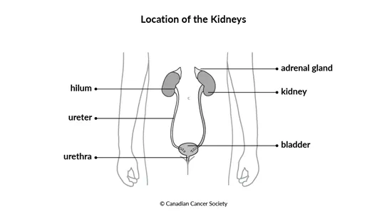
The kidney is part of the
During pregnancy, the kidneys are some of the first organs to develop in a fetus. Cells called nephroblasts grow and divide quickly to create the kidneys. At a certain point, the nephroblasts stop growing and dividing and they mature into kidney cells. But sometimes nephroblasts continue to grow out of control instead of maturing into kidney cells. These abnormal nephroblasts may lead to non-cancerous (benign) tumours such as congenital mesoblastic nephroma, which develops in newborns and infants.
In some cases, the abnormal nephroblasts can cause
Wilms tumour is the most common type of kidney cancer in children. Itʼs usually diagnosed in children 2 to 4 years old.
Most Wilms tumours are found in only one kidney (called unilateral Wilms tumour). In
about 5% of cases, it is found in both kidneys (called bilateral Wilms tumour). It
may be in both kidneys at the time of diagnosis, or it may develop in the other
kidney a few years after being found in the first kidney. Bilateral Wilms tumour is
more common in children who have an
Types of Wilms tumour
There are 2 types of Wilms tumour, and each type is determined by how the cells
look under a microscope (called histology). Doctors tell the 2 types apart based
on whether anaplasia is present. Anaplasia is a loss of
Favourable histology
About 90% to 95% of Wilms tumours are classified as having a favourable histology, which means there is no anaplasia present.
Wilms tumours with a favourable histology respond better to chemotherapy, so they have a better prognosis (outcome) than tumours with anaplasia present (anaplastic histology).
Anaplastic histology
About 5% to 10% of Wilms tumours are classified as having an anaplastic histology. An anaplastic histology may also be called an unfavourable histology. The cells in these tumours look very different from normal kidney cells and divide abnormally.
Anaplasia may be focal or diffuse.
Focal anaplasia means anaplasia is found only in one or a few specific areas of the tumour. The rest of the tumour looks like a regular (favourable histology) Wilms tumour.
Diffuse anaplasia means anaplasia is spread throughout the tumour.
Anaplastic tumours are more difficult to treat because they tend to be more resistant to chemotherapy. Anaplastic tumours are more likely to recur (come back) after treatment than tumours with a favourable histology.
The kidneys
Other childhood kidney tumours
Your trusted source for accurate cancer information
With support from readers like you, we can continue to provide the highest quality cancer information for over 100 types of cancer.
We’re here to ensure easy access to accurate cancer information for you and the millions of people who visit this website every year. But we can’t do it alone.
Every donation helps fund reliable cancer information, compassionate support services and the most promising research. Please give today because every contribution counts. Thank you.