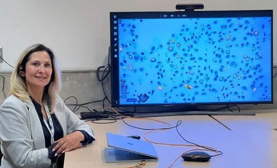
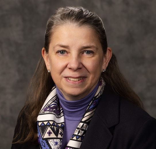
Dr April Khademi makes no promises when it comes to using artificial intelligence (AI) for medical imaging. She doesn’t need to. After designing algorithms for medical images for more than a decade, the results of her AI-powered research speak for themself.
“Humans can make mistakes,” she says. “But AI tools can help us to be more accurate and consistent.”

A biomedical engineer at the Toronto Metropolitan University, Dr Khademi is funded by a CCS Emerging Scholar Award. She is using this funding to create an AI tool that helps pathologists make more consistent and reliable breast cancer diagnoses that are used to make treatment decisions. Her team’s recent Scientific Reports publication highlights AI’s role in boosting accuracy, optimizing workflow and ultimately enhancing patient care and improving outcomes.
As part of their CCS-funded research, Dr Khademi’s team tested the AI tool they created with 90 pathologists around the world. Dr Susan Done, a pathologist and researcher at Princess Margaret Cancer Centre in Toronto, provided input into the study design and analysis of the data.
“The potential is significant,” she says. “The goal of AI is to make the assessment more standardized and more reproducible. If I assessed a slide using AI and my colleague across the street assessed the same slide next week, we’ll get the same score. That’s standardization.”
Dr Done says standardization will enable the testing of specific tumour markers, such as the breast marker targeted in Dr Khademi’s project, to be incorporated into routine clinical care.

How Dr Khademi's work is helping change outcomes
Currently, cancer is diagnosed by a pathologist, a specialized doctor who analyzes tissue samples to identify cancer presence and type.
“Pathology is the definitive diagnosis for cancer,” says Dr Khademi, the Canada Research Chair in AI for Medical Imaging. “Traditionally, it’s performed using a microscope to look at cells and tissues and provide scores and grades that are used to decide treatments.”
This poses a challenge because different pathologists may interpret the same sample differently. This is because they use qualitative descriptors such as colour and shape to make their assessment. Even when using size as an indicator, there can be variations on where measurements are taken. And with a shortage of pathologists, this method of diagnosing cancer and determining treatment can lead to delays in diagnosis and care.
“With any human-based analysis, depending on your experience, training or even the time of the day, you might get different answers when visually interpreting an image,” Dr Khademi says. “AI, on the other hand, can provide quantitative measurements and reduce variability in data for improved quality of care.
”Amanda Dy, a PhD student on Dr Khademi’s team, has seen firsthand how the tool makes pathologists more accurate and creates consensus among colleagues.
“It also helps them work faster to improve diagnostic turnaround time,” she adds.
Dr Khademi’s team is now working to apply the tool more widely on newer and bigger sets of data to further test reliability before it becomes standard practice.
CCS funding has given me the opportunity to make AI medical imaging more robust and I can’t thank the donors enough for their support. I would like to see pathologists using our tool one day so we can ultimately improve patient care.
To learn more about how CCS is investing in cutting-edge research, check out our research strategy.
