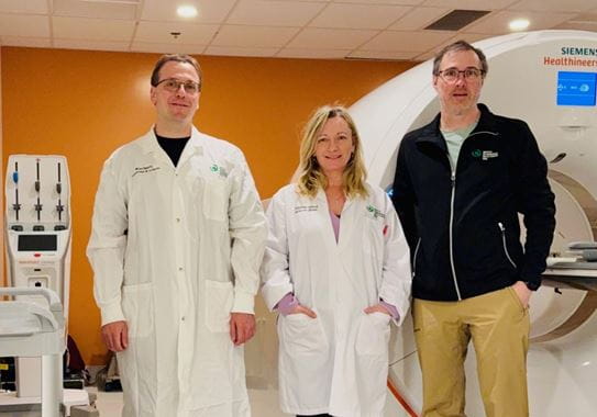
Getting accurate images of breast cancer is essential for choosing the right treatment, especially for cancers caused by estrogen. That’s why Dr Eric Turcotte at the Université de Sherbrooke is leading a CCS-funded research team to test a new imaging method that shows these tumours more clearly than current scans.
“Breast cancer can be hard to find on regular imaging,” says Dr Turcotte. “So we wanted to develop a new test targeting estrogen receptors. Now we’re scanning from head to toe – anywhere the cancer can hide, we’re able to see it.”
The imaging technique, called 4FMFES-PET, uses a special tracer liquid that is injected into the bloodstream. When the tracer finds a cell expressing estrogen receptors, it stays there.
Lobular breast cancer is a type of cancer that often has high numbers of estrogen receptors. Early results show that 4FMFES-PET imaging works especially well for this cancer, identifying a patient group that would most benefit from this new approach. The scans are proving to be both accurate and sensitive, finding cancer even when older methods cannot.
“For this kind of cancer, it is one of the best tracers in the world,” says Dr Michel Paquette, a research associate in Dr Turcotte’s lab. “We try our best to make it available to people. It’s a potential game changer.”
Supported by a CCS Innovation to Impact grant, the research team launched a clinical trial to explore the new technique’s full potential. The study began in Quebec, but now also includes people in Ontario.
Joanne Dubreuil, a 67-year-old former nurse, was recently diagnosed with breast cancer. When she heard about Dr Turcotte’s trial, she decided she wanted to join.
“As long as you have cancer, it would be nice if it served a purpose,” she says. “You have to make yourself useful with it.”
Surgeons successfully removed Joanne’s tumour, but the trial helped confirm that there were no traces of cancer left in her body.
“I was happy to take part in research,” she says. “At the same time, I got a new scan that could more precisely determine whether there were tumours elsewhere.”
Dr Turcotte’s team was pleased to confirm that the cancer had been fully removed.
If the research continues to show strong results, 4FMFES-PET could enhance or even replace current scanning methods to help people make more informed decisions about cancer treatment. The team emphasizes how crucial CCS funding has been for this clinical trial.
“CCS funding was the major pillar supporting everything,” Dr Turcotte says. “Without it, we wouldn’t have been able to scan and help as many patients.”

I was happy to take part in research. At the same time, I got a new scan that could more precisely determine whether there were tumours elsewhere.
Joanne Dubreuil, breast cancer survivor and trial participant
Help create a future without cancer
With support from readers like you, we can continue to make a meaningful impact for people affected by cancer.
We are determined to increase survival, stop cancer before it starts, and improve lives. But we can’t do it without you.
If everyone reading this gave just $5, we could achieve our goal this month to fund the most promising research, compassionate support and transformative advocacy. Please give today because every contribution counts. Thank you.